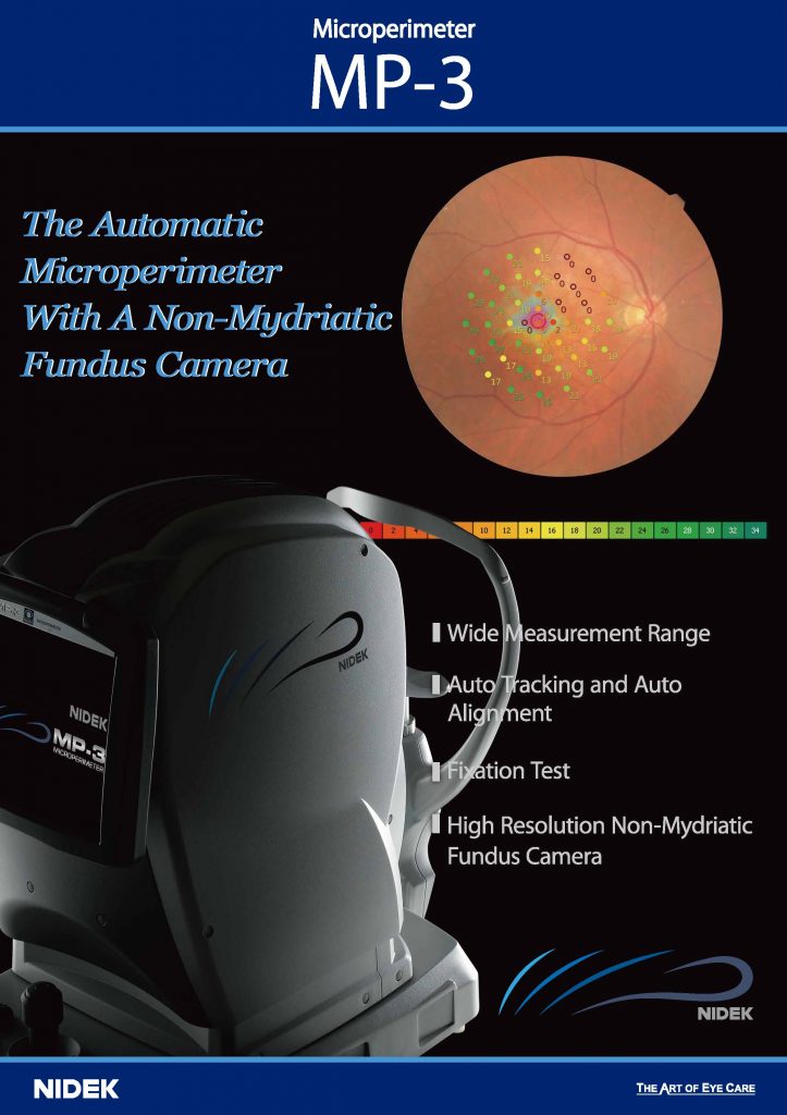
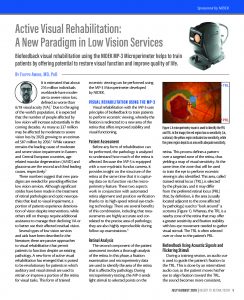
Active Visual Rehabilitation: A New Paradigm in Low Vision Services
Biofeedback visual rehabilitation using the NIDEK MP-3 Microperimeter helps to train patients by offering potential to restore visual function and improve quality of life.
By Filippo Amore, MD, PhD
It is estimated that about 216 million individuals worldwide have moderate to severe vision loss, defined as worse than 6/18 visual acuity (VA).1 Due to the aging of the world’s population, it is expected that the number of people affected by low vision will increase substantially in the coming decades. As many as 337 million may be affected by moderate to severe vision loss by 2020, growing to an estimated 587 million by 2050.1 While cataract remains the leading cause of moderate and severe vision impairment in Eastern and Central European countries, age-related macular degeneration (AMD) and glaucoma are the second and third leading causes, respectively.
These numbers suggest that new paradigms are needed for providing effective low vision services. Although significant strides have been made in the treatment of retinal pathologies and optic neuropathies that lead to visual impairment, a portion of patients experience deterioration of vision despite interventions, while others still on therapy require additional assistance to manage their declining VA or to better use their affected residual vision.
Several types of low vision services and aids have been described in the literature; these are passive approaches to visual rehabilitation that permit patients to function despite an existing pathology. A new form of active visual rehabilitation has emerged that is poised to be revolutionary for patients in which auditory and visual stimuli are used to retrain or improve a portion of the retina for visual tasks. This form of trained eccentric viewing can be performed using the MP-3 Microperimeter developed by NIDEK.
Filippo Amore, MD, PhD
Director of the National Center of Services for the Prevention of Blindness and Rehabilitation of the Visually Impaired, Rome, Italy
VISUAL REHABILITATION USING THE MP-3
Visual rehabilitation with the MP-3 uses principles of biofeedback to train patients to perform eccentric viewing, whereby the fixation is redirected to a new area of the retina that offers improved stability and visual functioning.
Patient Assessment
Before any form of rehabilitation can be performed, the pathology is analyzed to understand how much of the retina is affected. Because the MP-3 is equipped with a non-mydriatic fundus camera, it provides insight on the structure of the retina at the same time that it is capturing data on its function via the microperimetry feature. These two aspects work in conjunction with automated retina alignment and position verification thanks to its high-speed retinal eye-tracking technology. There are several benefits of this combination, including that measurements are highly accurate and correlated to the precise area of pathology; they are also highly reproducible during follow-up examinations.3
Retinal Analysis
The second component of the patient assessment involves a thorough analysis of the retina. In this phase, a fixation examination and microperimetry data are used to identify the area of the retina that is affected by pathology. During microperimetry testing, the MP-3 sends light stimuli to selected points on the retina. This process defines a pattern over a targeted zone of the retina, thus yielding a map of visual sensitivity. At the same time, the zone that will be used to train the eye to perform eccentric viewing is also identified. This area, called trained retinal locus (TRL), is selected by the physician, and it may differ from the preferred retinal locus (PRL) that, by definition, is the area (usually located adjacent to the zone affected by pathology) used to “look around” a scotoma (Figure 1). Whereas, the TRL is a nearby zone of the retina that may offer a greater sensitivity and fixation stability with less eye movement needed to gather visual stimuli. The TRL is often selected over or close to the patient’s PRL.
Biofeedback Using Acoustic Signals and Flickering Stimuli
During a training session, an audio cue is used to guide the patient’s fixation to the TRL. This is done by an intermittent audio cue; as the patient moves his/her eye to align fixation toward the TRL, the sound becomes more consistent,culminating in a continuous sound once fixation on the TRL is achieved. Then, to reinforce stability on the new area, a flickering checkerboard (Figure 2) is projected on the TRL. This checkerboard can be adapted to the needs of the patient in terms of size and flickering speed. Our research has suggested that optimal results are achieved with a minimum of 10- to 15-minute training sessions, twice a week, for 5 to 7 weeks, reaching a total of 10 to 15 training sessions.